

The National Institute of Statistical Sciences (NISS) and Merck sponsored a Virtual Meet-Up on the use of Statistical and Deep Learning Methods for Biomedical Images on Tuesday, March 15, 2022 from 11 am to 12:30 pm ET.
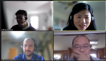
This meetup provided a snapshot of this vital interdisciplinary area with three talks, delivered by speakers from academic and industrial backgrounds. The speakers for this Virtual Meet-Up included Dr. John Kang from Merck, Dr. Hongtu Zhu from the University of North Carolina, and Dr. Andrew Janowczyk, from Case Western Reserve University. The moderator for this session was Dr. Peining Tao from Merck.
The presenters explained how imaging plays an important role in clinical diagnosis, biomedical research, and pharmaceutical research & development. For decades, innovative statistical methods have been applied to various biomedical image modalities. In the past 10 years, deep learning and AI have increasingly dominated this field. Recently, biomedical image analysis has become an extremely complex area, because of the diversity both in image modality and the analysis methodology.
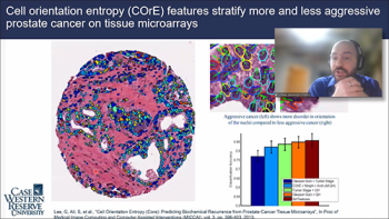
Andrew Janowczyk was our first speaker for the meetup and presented on “Computational Pathology: Towards Precision Medicine.” He gave the audience a very informative introduction to Computational Digital Pathology, including the Research Applications and talked about HistoSuite Tools.
Andrew highlighted reasons why Computer Aided Diagnostics (CAD) is useful and explained how the vast amounts of data could be used efficiently. He clarified the differences between Research and Clinical CAD Applications and elaborated about deep learning methods which are much faster than creating hand-crafted features. Whereas hand-crafted nuclei segmentation has taken three years, deep learning takes three hours. He next provided a few impressive examples of research applications. One example was the Nuclear Shape and Orientation Features from H&E Images to predict survival in Early Stage Estrogen Receptor Positive (ER+) Breast Cancers.
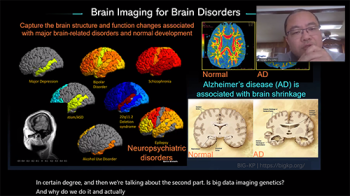
Our next speaker Hongtu Zhu, speaking on “Biobank-scale Multi-organ Imaging Genetics and Beyond,” provided specific examples of methodological challenges, big data in imaging genetics, and highlighted some novel clinical findings. These included examples of large-scale medical studies, brain imaging modality, and he also highlighted the methodological challenges of imaging genetics. Hongtu explained that these examples are “just a beginning” and that there are actually hundreds of associated genetic variants for 1593+ neuroimaging traits across various modalities: (grey matter volume, white matter microstructure, resting-state functional connectivity+rfMRI, task fMRI, shape, heart).
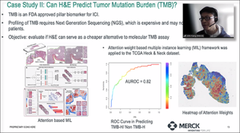
Our final speaker John Kang presented on “Characterizing Tumor Micro Environment (TME) Using H&E Images” which gave the background of patho-mics/digital pathology in translational oncology. He provided an overview of the internal analytic workflow, provided examples from case studies and outlined the future direction of deep learning methods for biomedical images at Merck.
While H&E Images are commonly used in path-omics analyses, and provide detailed representation of tumor architecture with high levels of cytologic, morphologic, and structural detail, John explained that H&E images still suffer from some major limitations, so challenges still exist to focus on for the future.
This was a very enlightening NISS-Merck Meetup and we thank all the speakers and the Merck organizer, Junshui Ma, for making this an exceptional event.
The full recording of this event is available on our NISS YouTube Channel below.
Recording of the Session
Slides Used by the Speakers
Dr. Andrew Janowczyk, (Case Western Reserve University)
"Computational Pathology: Towards Precision Medicine"
Dr. John Kang, (Merck)
"Characterizing Tumor Micro Environment (TME) Using H&E Images"
Dr. Hongtu Zhu, University of North Carolina
